540 εκατ χρόνια πρινΕξελίσσονται τα πρώτα Εντερόπνευστα ζώα, δηλαδή σκουλήκια, τα οποία είναι οι πρώτοι οργανισμοί που έχουν καρδιά και και νεφρό και διαθέτουν το πρώτο σύστημα βράγχιων
 http://www.tovima.gr/science/medicine-b ... aid=503068
Ιδού το σκουλήκι-«φαλλός»
Τα απολιθώματά του βρέθηκαν στον Καναδά και δείχνουν τον «χαμένο κρίκο» της εξέλιξης κάποιων θαλάσσιων όντων
Λονδίνο
Ένας προϊστορικός σκώληκας με σχήμα φαλλού φαίνεται ότι αποτελεί τον σημαντικό «χαμένο κρίκο» της εξέλιξης ορισμένων θαλάσσιων όντων. Απολιθώματα αυτού του… μακρουλού όντος με την επιστημονική ονομασία Spartobranchus tenuis ήλθαν στο φως στο Εθνικό Πάρκο του Γιόχο στον Καναδά το οποίο είναι διάσημο για τα απολιθώματα της Κάμβριας Περιόδου που κρύβει – πρόκειται για μια περίοδο που ξεκίνησε πριν από 542 εκατομμύρια χρόνια και κατά την οποία υπήρξε «έκρηξη» της βιοποικιλότητας στη Γη.
Σε περιοχή ηλικίας 505 εκατομμυρίων ετών
Ο χαμένος κρίκος της θαλάσσιας εξέλιξης εντοπίστηκε από ερευνητές του Πανεπιστημίου του Μόντρεαλ. Όπως αναφέρουν οι ειδικοί στην επιθεώρηση «Nature», τα απολιθώματα του «φαλλικού» Spartobranchus tenuis εντοπίστηκαν σε μια περιοχή η οποία εκτιμάται ότι είναι περίπου 505 εκατομμυρίων ετών.
Το περίεργο στην όψη ασπόνδυλο ον θεωρείται ως ένας άγνωστος πρόγονος των εντερόπνευστων, σπάνιων επιμηκών σκωλήκων με μήκος από λίγα εκατοστά μέχρι δύο μέτρα, τα οποία ζουν μέσα σε στοές σε ιζήματα του βυθού ή κάτω από πέτρες. Τα εντερόπνευστα διαθέτουν προβοσκίδα επάνω στην οποία φέρουν βλεφαριδωτές ταινίες που εξυπηρετούν τη διατροφή και τη μετακίνησή τους.
Όπως εξήγησε ο επικεφαλής της μελέτης δρ Κρις Κάμερον «σε αντίθεση με άλλα ζώα που διέθεταν σκληρές επιφάνειες όπως δόντια, οστά ή λέπια, αυτού του είδους οι σκώληκες είχαν πολύ μαλακό σώμα με αποτέλεσμα το αρχείο απολιθωμάτων τους να είναι εξαιρετικά σπάνιο».Ο ερευνητής προσέθεσε ότι «η περιγραφή του Spartobranchus tenuis που κάνουμε σε αυτή τη δημοσίευση, ενός όντος άγνωστου μέχρι σήμερα στην επιστήμη, πηγαίνει το αρχείο απολιθωμάτων των εντερόπνευστων 200 εκατομμύρια χρόνια πίσω, στην Κάμβρια περίοδο, αλλάζοντας εκ βάθρων τη γνώση μας σχετικά με τη βιοποικιλότητα εκείνη την εποχή».
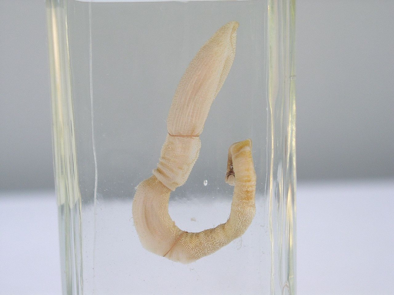
Oι σημερινοί απόγονοι του σκώληκα-φαλλού ανήκουν στα εντερόπνευστα, σπάνια επιμήκη σκουλήκια με μήκος από λίγα εκατοστά μέχρι δύο μέτρα, τα οποία ζουν μέσα σε στοές σε ιζήματα του βυθού ή κάτω από πέτρες (Credit:C.B. Cameron)
Η ανατομία του… μακρουλού σκώληκα
Η ενδελεχής ανάλυση των απολιθωμάτων έδειξε ότι το σκουλήκι… πέος διέθετε ένα ευλύγιστο σώμα το οποίο αποτελείτο από μια κοντή προβοσκίδα, ένα κολάρο και έναν στενό επιμήκη κορμό ο οποίος κατέληγε σε μια σφαιρική δομή που πιθανότατα είχε τον ρόλο «άγκυρας».
Τα μεγαλύτερα δείγματα που ανέσυραν οι επιστήμονες είχαν μήκος 10 εκατοστών εκ των οποίων μισό εκατοστό ήταν το μήκος της προβοσκίδας. Οι ερευνητές εκτιμούν ότι τα σκουλήκια αυτά τρέφονταν με μικρά σωματίδια τα οποία φίλτραραν από το νερό της θάλασσας.
Ο χαμένος «κρίκος»
Οι σύγχρονοι απόγονοι του Spartobranchus tenuis, τα εντερόπνευστα, αποτελούν μέλη των ημιχορδωτών, μιας ομάδας θαλάσσιων ζώων που συνδέονται στενά με τους σημερινούς αστερίες και τους αχινούς (ανήκουν στα πτεροβράγχια). Από την ανακάλυψή τους τον 19ο αιώνα υπήρχαν πολλά και σημαντικά ερωτήματα σχετικά με την εξέλιξη των ημιχορδωτών – μεταξύ άλλων στο σκοτάδι παρέμενε η σχέση μεταξύ εντρόπνευστων και πτεροβράγχιων.
Όπως φαίνεται τελικώς τα εντερόπνευστα και τα πτεροβράγχια μοιάζουν πολύ διαφορετικά, ωστόσο μοιράζονται πολλά γενετικά και αναπτυξιακά χαρακτηριστικά.
Οι ερευνητές σημειώνουν ότι οι καινούργιες αναλύσεις σχετικά με τον Spartobranchus tenuis δείχνουν ότι ο σκώληκας αυτός αποτέλεσε τον κοινό πρόγονο των δύο ομάδων παρέχοντας τελικά στους επιστήμονες τον χαμένο εξελικτικό κρίκο που αναζητούσαν.
Ο σημαντικός «χαμένος κρίκος»: ο προϊστορικός σκώληκας με το περίεργο σχήμα θεωρείται ότι αποτελεί τον άγνωστο μέχρι σήμερα πρόγονο των εντερόπνευστων καθώς και των αστεριών και των αχινών που είναι γνωστά ως πτεροβράγχια (Credit:Marianne Collins)


Structure of branchial region – bc, coelom. tb, tongue-bars. ds, mesentery. pr, ridge. vv, vessel. gp, gill-pore. dn, dorsal nerve. dv, vessel. œ, oesophagus. vs, mesentery. vn, ventral nerve.
Digestive system[edit]
Many acorn worms are detritus feeders, eating sand or mud and extracting organic detritus. Others feed on organic material suspended in the water, which they can draw into the mouth using the cilia on the gill bars.[13] A groove lined with cilia lies just in front of the mouth and directs suspended food into the mouth and may allow the animal to taste.[12]
The mouth cavity is tubular, with a narrow diverticulum or stomochord extending up into the proboscis. This diverticulum was once thought to be homologous with the notochord of chordates, hence the name "hemichordate" for the phylum. The mouth opens posteriorly into a pharynx with a row of gill slits along either side. The remainder of the digestive system consists of an oesophagus and intestine; there is no stomach.[12]
In some families there are openings in the dorsal surface of the oesophagus connecting to the external surface, through which water from the food can be squeezed, helping to concentrate it. Digestion occurs in the intestine, with food material being pulled through by cilia, rather than by muscular action.[12]
Acorn worms breathe by drawing in oxygenated water through their mouth. The water then flows out the animal's gills which are on its trunk. Thus, the acorn worm breathes about the same way as fish.
Circulatory system[edit]
Acorn worms have an open circulatory system, in which the blood flows through the tissues sinuses. A dorsal blood vessel in the mesentery above the gut and delivers blood to a sinus in the proboscis that contains a muscular sac acting as a heart. Unlike the hearts of most other animals, however, this structure is a closed fluid-filled vesicle whose interior does not connect directly to the blood system. Nonetheless, it does regularly pulsate, helping to push blood through the surrounding sinuses.[12]
From the central sinus in the collar, blood flows to a complex series of sinuses and peritoneal folds in the proboscis. This set of structures is referred to as a glomerulus and may have an excretory function, since acorn worms otherwise have no defined excretory system. From the proboscis, blood flows into a single blood vessel running underneath the digestive tract, from which smaller sinuses supply blood to the trunk, and back into the dorsal vessel.[12]
The blood of acorn worms is colourless and acellular.[12]
Respiratory system[edit]
Acorn worms continually form new gill slits as they grow in size, with some older individuals having more than a hundred on each side. Each slit consists of a branchial chamber opening to the pharynx through a U-shaped cleft and to the exterior through a dorso-lateral pore (see diagram below). Cilia push water through the slits, maintaining a constant flow. The tissues surrounding the slits are well supplied with blood sinuses.[12]
Nervous system[edit]
A plexus of nerves lies underneath the skin, and is concentrated into both dorsal and ventral nerve cords. While the ventral cord runs only as far as the collar, the dorsal cord reaches into the proboscis, and is partially separated from the epidermis in that region. This part of the dorsal nerve cord is often hollow, and may well be homologous with the brain of vertebrates. In acorn worms, it seems to be primarily involved with coordinating muscular action of the body during burrowing and crawling.[12]
Acorn worms have no eyes, ears or other special sense organs, except for the ciliary organ in front of the mouth, which appears to be involved in filter feeding and perhaps taste (3). There are, however, numerous nerve endings throughout the skin.[12]
Similarities to chordates[edit]
Acorn worms are considered more highly specialised and advanced than other similarly shaped worm-like creatures. They have a circulatory system with a heart that also functions as a kidney.[citation needed] Acorn worms have gill-like structures that they use for breathing, similar to the gills of primitive fish. Therefore, acorn worms are sometimes said to be a link between classical invertebrates and vertebrates. Some also have a postanal tail which may be homologous to the post-anal tail of vertebrates. An interesting trait is that its three-section body plan is no longer present in the vertebrates, except for the anatomy of the frontal neural tube, later developed into a brain which is divided into three main parts. This means some of the original anatomy of the early chordate ancestors is still present even if it is not always visible.
One theory is that the three-part body originates from an early common ancestor of all the deuterostomes, and maybe even from a common bilateral ancestor of both the deuterostomes and protostomes.[citation needed] Studies have shown that the gene expression in the embryo share three of the same signaling centers that shape the brains of all vertebrates, but instead of taking part in the formation of their neural system,[14] they are controlling the development of the different body regions.

Structure of anterior end – a, Arrow from proboscis-cavity (pc) passing to left of pericardium (per) and out through proboscis pore-canal. b1, arrow from central canal of neurochord (cnc) passed out through anterior neuropore. b2, ditto; through posterior neuropore. c, arrow intended to pass from 1st gill-pouch through collar pore-canal into collar-coelom (cc). cts, posterior limit of collar. dv, dorsal vessel passing into central sinus (bs). ev, efferent vessel passing into ventral vessel (vv). epr, epiphysial tubes. st, stomochord. vs, ventral septum of proboscis. sk, body of nuchal skeleton. m, mouth. th, throat. tb, tongue-bars. tc, trunk coelom.